

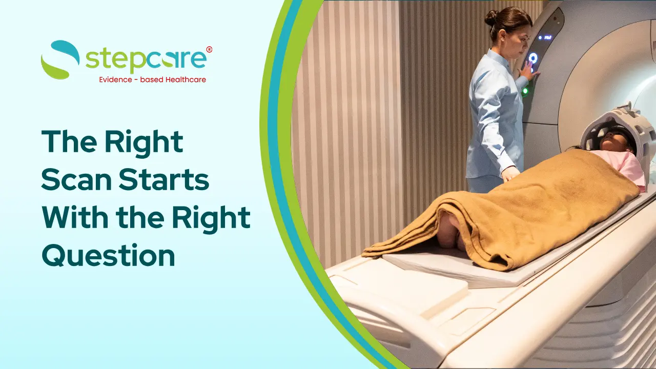
MRI vs Ultrasound for Joint and Back Pain: Which Scan Do You Need in Whitefield?
If your doctor has mentioned a scan for your joint or back pain and you are trying to understand the difference between MRI and ultrasound, the choice is not about which is better. It is about which one answers the clinical question you have.
Here is the short answer: ultrasound is excellent for superficial soft tissue structures like tendons, bursae, and superficial muscle tears, and it allows dynamic assessment while you move. MRI is the definitive investigation for deep structures, spinal discs, nerve compression, bone marrow, and internal joint structures like menisci and cruciate ligaments.
How Each Scan Works
Ultrasound uses high-frequency sound waves. A probe is moved over the skin, and the reflected sound waves build a real-time image. It is radiation-free, painless, inexpensive, and can be done while you move the joint.
MRI uses a strong magnetic field and radio waves to produce highly detailed cross-sectional images of both soft tissue and bone. It is radiation-free but requires you to lie still inside an enclosed scanner for 20 to 45 minutes.
X-ray, worth mentioning alongside, uses ionising radiation and is the first-line test for bony problems: fractures, joint space narrowing in arthritis, and spinal alignment.
MRI vs Ultrasound: A Direct Comparison
|
|
|
|
|
|
|
|
|
|
|
|
|
|
|
|
|
|
|
|
|
|
|
|
|
|
|
|
|
|
|
|
|
|
|
|
|
|
|
|
|
|
|
|
|
|
|
|
When Ultrasound Is the Right Choice
Ultrasound is the appropriate investigation for:
- Shoulder: rotator cuff tendinopathy and partial tears, subacromial bursitis, biceps tendon problems
- Elbow: tennis elbow (lateral epicondylitis), golfer's elbow
- Wrist and hand: de Quervain's tenosynovitis, ganglion cysts, trigger finger
- Knee: Baker's cyst, patellar tendinopathy, superficial bursitis, joint effusion
- Ankle and foot: Achilles tendinopathy, plantar fasciitis, ankle ligament assessment
- Muscle injuries: hamstring and calf tears
- Guided procedures: injections into joints, bursae, or tendon sheaths under real-time visualisation
The dynamic advantage matters clinically. Some shoulder impingement problems and tendon subluxations are only visible when the joint is moving, which MRI cannot capture.
When MRI Is the Right Choice
MRI is necessary for:
- Back and neck pain with radiation into the limbs, suggesting disc prolapse or nerve root compression
- Suspected spinal canal stenosis
- Knee internal derangement: meniscal tears, ACL and PCL injuries
- Suspected stress fracture where X-ray is normal but pain persists
- Bone marrow oedema, avascular necrosis, or bone infection
- Labral tears in the shoulder or hip
- Cartilage assessment
- Suspected tumour or infection in bone or deep soft tissue
- Pre-surgical planning for orthopaedic procedures
If your back pain radiates down the leg with numbness, tingling, or weakness, that is a nerve involvement picture, and MRI is the correct investigation. Ultrasound cannot assess the spine.
Do You Need Any Scan at All?
This is worth asking. Most acute back pain and many joint pains resolve with conservative management within 4 to 6 weeks and do not require imaging at all. International guidelines specifically advise against routine imaging for uncomplicated back pain in the first 6 weeks.
Imaging is indicated when: symptoms persist beyond 6 weeks despite appropriate treatment, there are red flag features such as significant trauma, unexplained weight loss, fever, or neurological deficit, a specific structural diagnosis would change management, or surgery is being considered.
Scanning too early often reveals incidental age-related changes that are present in many symptom-free people and can lead to unnecessary anxiety and intervention.
The Practical Sequence Most Doctors Follow
|
|
|
|
|
|
|
|
|
|
|
|
|
|
|
|
|
|
|
|
Book an Ultrasound or Imaging Consultation at Stepcare Whitefield
Stepcare offers musculoskeletal ultrasound and digital X-ray in-house, with physician consultation to determine which investigation your symptoms actually warrant before you spend on a scan.
Call or WhatsApp: +91 9606910113 Email: appointments@stepcare.co.in Address: G-01, Ground Floor, Brigade IRV Center, Nallurhalli, Whitefield, Bengaluru 560066 Hours: Monday to Saturday, 8 AM to 8 PM | Sunday, 10 AM to 6 PM
Disclaimer: This content is for informational purposes only and should not be considered medical advice. Please consult a qualified healthcare professional before making any medical decisions.
Need care guidance?
Book a consultation with Stepcare and get professional support for your health concern.
Book Appointment